
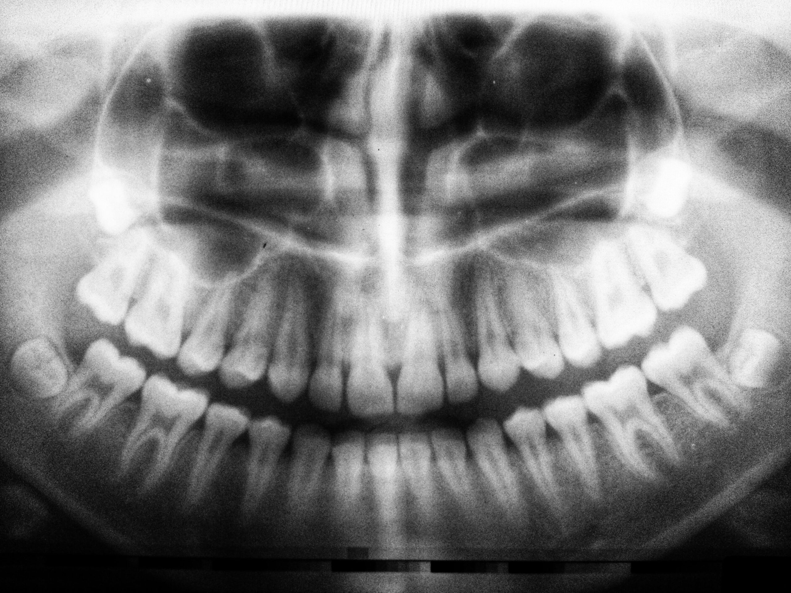
What are dental X-rays?
Dental X-rays ,also known as radiographs, are internal images of your teeth and jaws.They are part of the routine dental examination and help the dentist detect hidden problems . These could be tooth decay,low bone levels,root infection or buried roots.
Why are X-rays important?
Visual examination is not always sufficient to identify all of the problems in your mouth. Radiographs reveal conditions you may have not been aware of. Early detection of a problem will allow your dentist to diagnose and treat them before they become more serious.
How do the X-rays work?
While you seat in the dental chair your dentist will place special film or sensor in your mouth. They will then position X-ray machine tube over your face and press a button to capture the image.
What types of X-rays exist?
There are two main types of radiographs: intraoral and extraoral.
Intraoral: sensor or film is inside the mouth.
These could be bite wings,periapical or occlusal x-rays.They show different areas of the teeth.
Extraoral: sensor or film are outside your mouth
These are panoramic,cephalometric X-rays and CBCT scan. They show all of your teeth as well as jaws and skull structures.
Are dental X-rays safe?
Harmful effects from dental X-rays are very low because they give only a small amount of radiation. Digital X-rays in particular produce 80% less radiation than traditional ones. Radiographs though should not be taken more often than is necessary as in large doses they can increase cancer risk. It is also important for a female patient to advise their dentist if she is pregnant.
How often should I have X-rays?
Frequency of routine radiographs depends on the caries risk. The usual interval for a healthy mouth is 12-18 months. In case of recurring decay or periodontal disease though the interval should be reduced to 6 months.